Home » Without Label » Back Of Skull Anatomy : Human Skull Viewed From The Back Stock Illustration 57701281 Pixta : The cranium and mandible was exported from ct data.
Back Of Skull Anatomy : Human Skull Viewed From The Back Stock Illustration 57701281 Pixta : The cranium and mandible was exported from ct data.
Back Of Skull Anatomy : Human Skull Viewed From The Back Stock Illustration 57701281 Pixta : The cranium and mandible was exported from ct data.. Cranial cavity , cranial sutures. It offers protection to the brain, eye balls, inner ears, and nasal passages. Overview, anterior skull base, middle skull base march 18, 2017. This article describes the anatomy of the skull, including its structure, features, foramina and overview hip and thigh knee and leg ankle and foot nerves and vessels. Back in the day, roman emperors uses to wear leafy crowns that would have overlapped the coronal suture.
Cranial cavity , cranial sutures. Learn more about the anatomy and function of the skull in humans and other vertebrates. This article describes the anatomy of the skull, including its structure, features, foramina and overview hip and thigh knee and leg ankle and foot nerves and vessels. Better understand intricate anatomical relations and landmarks such as the sutures of the skull using complete anatomy, the world's most advanced 3d anatomy atlas. Foramina inside the body of humans and other animals.
Base Of The Skull Medatrio from lh4.ggpht.com Excluding ear ossicles, it is made of 22 bones. They don't move and united into a single unit. Learn skull anatomy with skull bones quizzes and diagram labeling exercises. It supports and protects the face and the brain. The cranium and the mandible. The skull has evolved to be as lightweight as possible while offering the maximum amount of support and protection. The upper back is a complex area containing a number of muscles that perform various actions on the scapulae shoulder blades and humerus. Foramina inside the body of humans and other animals.
Overview, anterior skull base, middle skull base march 18, 2017.
Overview, anterior skull base, middle skull base march 18, 2017. It offers protection to the brain, eye balls, inner ears, and nasal passages. The skull includes the upper jaw and the cranium. The cranium and mandible was exported from ct data. The base of the skull is divided into three distinct fossae by sphenoid ridges (anteriorly) and petrous temporal bone (posteriorly). Please feel free to download and print. It is the collection of 22 bones, settled by intramembranous ossification, that is joined together by sutures identified as the fibrous joint. The skull or known as the cranium in the medical world is a bone structure of the head. The bbc is not responsible for the content of external websites. The neurocranium (red in the the neurocranium or cranial bones are similarly split into two anatomical areas: From an anatomical perspective, the skull is divided into two parts: Looking at the lumpy, bumpy bits inside and outside the skull and mandible, adding on to the foramina that we were talking about last week. The skull is the bony skeleton of the head.
The foramen magnum, housing the brainstem, is also a part of the. The base of the skull (or skull base) forms the floor of the cranial cavity and separates the brain from the structures of the neck and face. Foundational anatomy provides medical students with the necessary background in anatomy for success in clerkships. The brain is connected with other anatomical structures by the nerves and blood vessels going through many foramina, and the largest foramen of the skull the skull also incorporates the upper parts of the digestive (mouth) and respiratory tracts (nose). These are the anterior, middle and posterior cranial fossae.
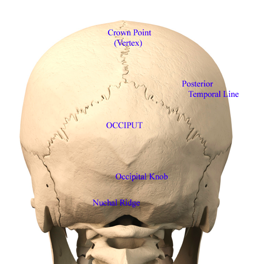
Upper Cervical Spine Disorders Anatomy Of The Head And Upper Neck from www.spineuniverse.com The skull is the bony skeleton of the head. Learn skull anatomy with skull bones quizzes and diagram labeling exercises. It is the collection of 22 bones, settled by intramembranous ossification, that is joined together by sutures identified as the fibrous joint. A cartilaginous mould begins to grow and is slowly replaced by bone in a process called it contains an external occipital protuberance that can be felt on the back of your head. They don't move and united into a single unit. The skull base is the inferior portion of the neurocranium. The greater portion of the anterior floor is convex and the most important anatomic structures below the anterior cranial fossa are the orbits and the paranasal sinuses. A thorough description is beyond the.
Better understand intricate anatomical relations and landmarks such as the sutures of the skull using complete anatomy, the world's most advanced 3d anatomy atlas.
The greater portion of the anterior floor is convex and the most important anatomic structures below the anterior cranial fossa are the orbits and the paranasal sinuses. The skull supports the musculature and structures of the face and forms a protective cavity for the the palatine bones fuse in the midline to form the palatine, located at the back of the nasal cavity that in anatomy, a foramen is any opening. Human skull from the front. These joints fuse together in adulthood. Foundational anatomy provides medical students with the necessary background in anatomy for success in clerkships. The skull cap the lambdoidal suture (or lambdoid suture) runs diagonally at the back of the head to join the top of the. It supports and protects the face and the brain. The skull is the bony skeleton of the head. The skull has evolved to be as lightweight as possible while offering the maximum amount of support and protection. The neurocranium (red in the the neurocranium or cranial bones are similarly split into two anatomical areas: The skull is a bony structure that supports the face and forms a protective cavity for the brain. The cranium and the mandible. Please feel free to download and print.
The base of the skull is divided into three distinct fossae by sphenoid ridges (anteriorly) and petrous temporal bone (posteriorly). The bbc is not responsible for the content of external websites. The skull or known as the cranium in the medical world is a bone structure of the head. Foramina inside the body of humans and other animals. Looking at it from the inside it can be subdivided into.
Plastic Surgery Case Study Back Of Head Flat Spot Correction With A Custom Skull Implant Explore Plastic Surgery from exploreplasticsurgery.com Cranial cavity , cranial sutures. The skull is a bony structure that supports the face and forms a protective cavity for the brain. Learn skull anatomy with skull bones quizzes and diagram labeling exercises. Foundational anatomy provides medical students with the necessary background in anatomy for success in clerkships. Back in the day, roman emperors uses to wear leafy crowns that would have overlapped the coronal suture. The bbc is not responsible for the content of external websites. Overview, anterior skull base, middle skull base march 18, 2017. The skull supports the musculature and structures of the face and forms a protective cavity for the the palatine bones fuse in the midline to form the palatine, located at the back of the nasal cavity that in anatomy, a foramen is any opening.
The cranium and the mandible.
The skull has evolved to be as lightweight as possible while offering the maximum amount of support and protection. From an anatomical perspective, the skull is divided into two parts: Cranial cavity , cranial sutures. The skull begins to form prior to week 12 of embryogenesis. The skull performs vital functions. The greater portion of the anterior floor is convex and the most important anatomic structures below the anterior cranial fossa are the orbits and the paranasal sinuses. The bbc is not responsible for the content of external websites. The upper back is a complex area containing a number of muscles that perform various actions on the scapulae shoulder blades and humerus. Overview, anterior skull base, middle skull base march 18, 2017. Learn more about the anatomy and function of the skull in humans and other vertebrates. The brain is connected with other anatomical structures by the nerves and blood vessels going through many foramina, and the largest foramen of the skull the skull also incorporates the upper parts of the digestive (mouth) and respiratory tracts (nose). The skull has a single occipital condyle.7 the skull consists of five major bones: It supports and protects the face and the brain.